Central optical works offers the highest quality of Zoology Models, Educational Zoology Models, Laboratory Zoology Models and botanical models. Custom Zoology Model, Zoology Models,Educational Zoology. Zoology Models,Zoological Models,Educational Zoology Models, Cheap Zoology Models,3D Zoological Models,Indian Zoology Models,Frog Dissection Models,Rat Dissection Models,Hydra Models,Earthworm Models

This model, enlarged about 20,000 times. Animal Cell Model, provides clear depiction of the delicate and complex structure of the animal cell. Shows the nucleus , endoplasmatic reticulum, mitochondria, ribosomes, polysomes and Golgi apparatus, centrioles, lysosomes and fat vacuoles.
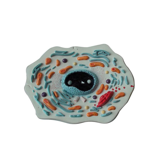
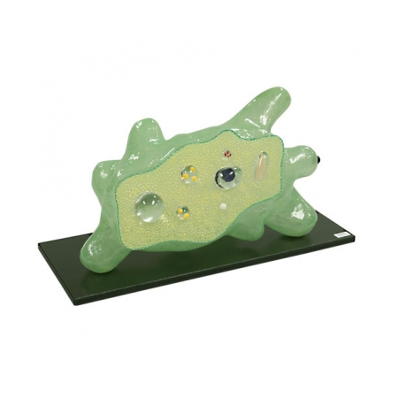
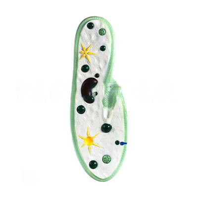
Animal Cell Three Dimensional Zoology Anatomy Model - A three dimensional model with one side dissected to show complete internal structure, mounted on base with key card.
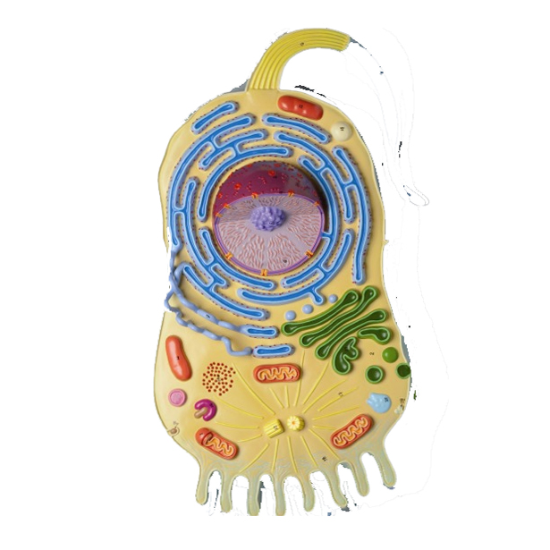
Model of cell showing ultrastructure, mounted on base with key card.
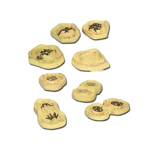
Robust Models illustrating the process of mitotic cell division. The chromosomes are painted to allow easy identification, mounted on base with key card.
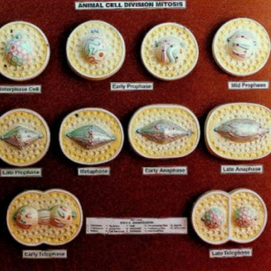
Animal Cell Division Mitosis - A set of 10 models, showing resting cell, early prophase, prophase, late prophase, metaphase, anaphase, late anaphase, Telophase and daughter cells, mounted on base with key card.
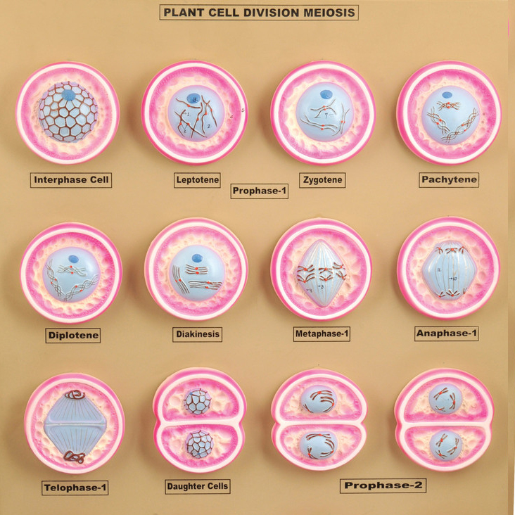
Animal Cell Division Meiosis Zoology Model - Set of 12 models, showing detailed stages in Meiosis cell division, beautifully coloured to bring out the detailed structure as seen under the microscope, mounted on base with key card.
Zoology Model Amoeba proteus, enlarged approx. 1000 times. In a small pseudopodium which can be opened up showing the structure after electron microscopic magnification, mounted on base with key card.
Paramecium, enlarged approx. 1600 times, showing the cell inventory of a protozoa: macro and micronucleus, contractile vacuoles, cytostome with membranellae, myonemes and food vacuoles and the formation of the endo and ectoplasm and the network of neuronemes, it also shows the structure of the pellicle of the ectoplasm, and the position and order of the trichocysts and a range of cilia in typical.
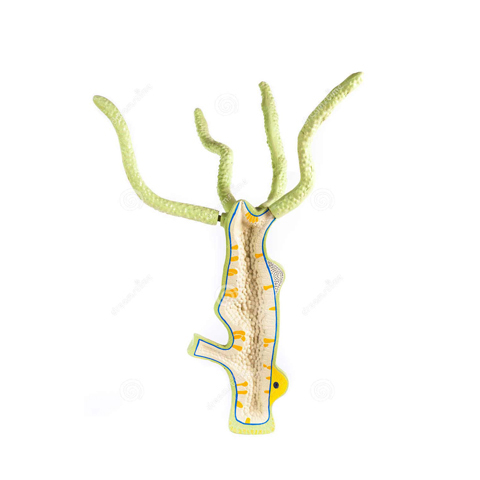
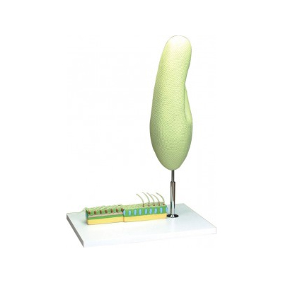
Model of Hydra - 3 Dimensional model dissection showing mouth coelentron, testis, gland cells, ovum, ovary etc., mounted on base with key card.
Tapeworm Zoology Model - Taenia solium, enlarged many times in one piece, mounted on base with key card.
DissectionModel.jpg)
Hen Dissection Model - Natural size, the right side shows the feathers and other side shows all the internal organs, mounted on base with key card.
Earthworm Dissection Model showing the external character in a portion and the dissection in the remaining, exhibiting all important systems, mounted on base with key card.
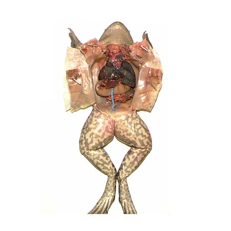
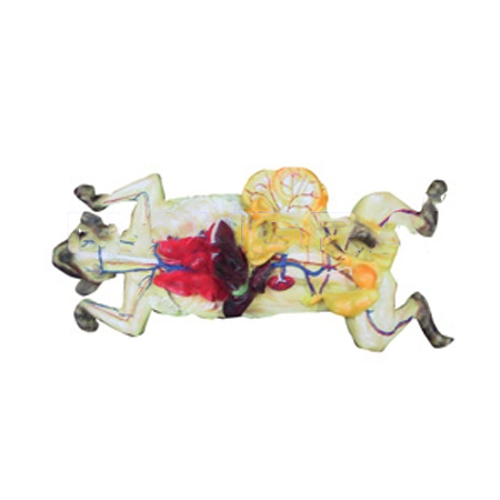
Model showing dissected structure of frog in detail, mounted on base with key card.
Perch Fish Dissection Model Showing all the general anatomy after dissection, mounted on base with key card.
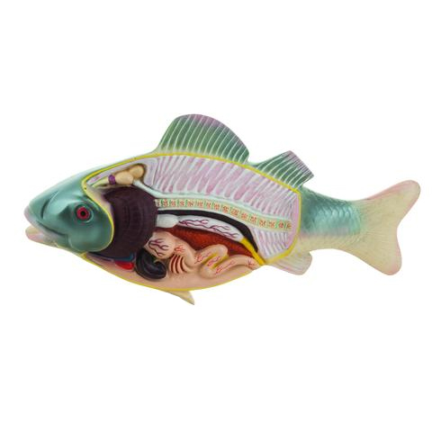
Fish Dissection Anatomy Model Showing all the general anatomy after dissection, mounted on base with key card.
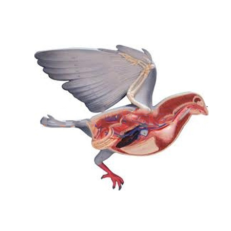
Pigeon Dissection - Bird Anatomy Zoology Model Natural size, dissection showing all the internal organs of a pigeon, mounted on base with key card.
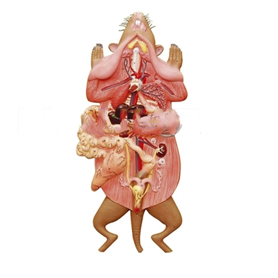
Rat Dissection Enlarged - Dissection model showing all the vital organs of a rat, mounted on base with key card.
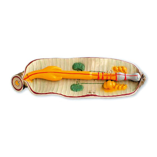
Paramecium Model Enlarged, dissection showing the structure, mounted on base with key card.
Enlarged, dissection showing the structure, mounted on base with key card.

Enlarged, dissection showing the internal organs of both male and female, mounted on base with key card.
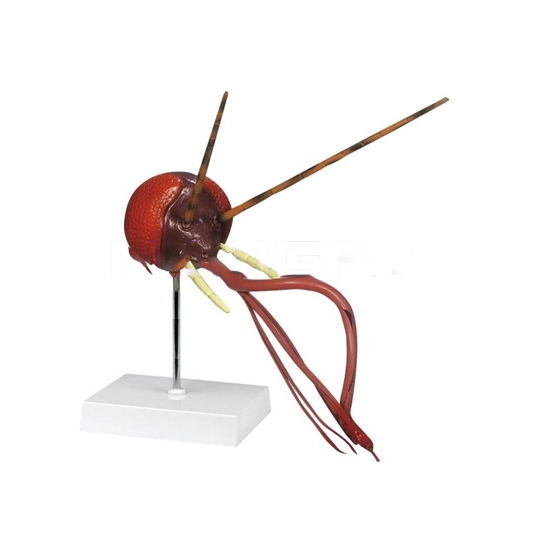
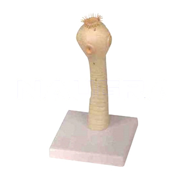
Model of Mosquito Head Showing different parts of mosquito head. Mounted on stand. Numbered with English Key Card.
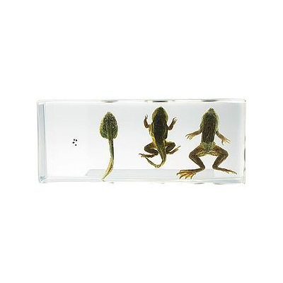
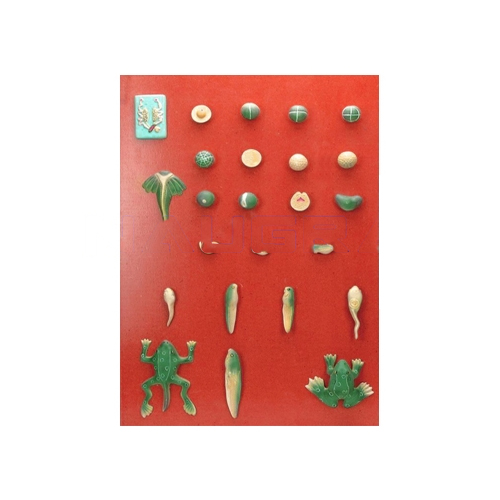
Frog Life Cycle Specimens - Frog Development specimens in presentation box showing different stages of the frog life cycle. Information card included. Specimens include egg, tadpole 6 weeks, tadpole with legs 9 weeks, tadpole with 4 limbs 10 weeks, tadpole with short tail 11 weeks, froglet and adult frog. These durable and clear specimens in lightweight acrylic provide students with a fascinating insight into animal life.
This life-size dissection model features over 100 individual anatomical details. An extremely realistic and precise model, it is crafted with hand-painted detail. Featured structures include a cross-sectioned kidney showing the cortex and medulla, major arteries and veins, muscle groups of the fore and hind limbs, and the open uterus exposing a developing fetus. This model also has an open mouth cavity detailing the teeth and nasopharynx, and includes a key identifying 136 structures.
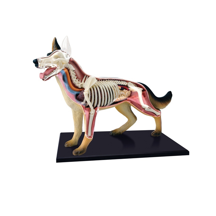
Dog Anatomy PVC life-size model features individual anatomical details. An extremely realistic and precise model, it is crafted with hand-painted detail.
Ambala Science Market © all rights reserved